

2024 Valid SPI Real Exam Questions, practice ARDMS SPI
Latest Success Metrics For Actual SPI Exam (Updated 110 Questions)
ARDMS SPI Exam Syllabus Topics:
| Topic | Details |
|---|---|
| Topic 1 |
|
| Topic 2 |
|
| Topic 3 |
|
| Topic 4 |
|
| Topic 5 |
|
NEW QUESTION # 10
Which factor does a string phantom evaluate?
- A. Intensity values
- B. Two-dimensional resolution
- C. Slice thickness
- D. Flow velocity
Answer: D
Explanation:
A string phantom is designed to evaluate the accuracy of Doppler ultrasound systems, specifically in measuring flow velocity. It consists of a moving string or filament that mimics blood flow within a vessel. By using this phantom, sonographers can assess how accurately the ultrasound system can detect and measure the speed of the moving target. This helps in calibrating and verifying the performance of Doppler systems, ensuring they provide accurate flow velocity readings in clinical practice.
Reference:
American Registry for Diagnostic Medical Sonography (ARDMS) Sonography Principles and Instrumentation study materials.
Textbook of Diagnostic Sonography by Hagen-Ansert, S. L. (latest edition).
NEW QUESTION # 11
Which adjustment is needed to optimize the waveform below?
- A. Increase pulse repetition frequency
- B. Decrease gain
- C. Lower baseline
- D. Increase wall filter
Answer: C
Explanation:
The waveform in the image shows spectral Doppler signals that are pushed against the upper limit of the display, indicating that the baseline is too high. Lowering the baseline allows for a better visual representation of the entire Doppler signal within the available display range. This adjustment prevents the waveform from being cut off and helps in accurately interpreting the blood flow characteristics.
Reference:
ARDMS Sonography Principles & Instrumentation Guidelines
Kremkau FW. Sonography Principles and Instruments. 9th ed. Philadelphia, PA: Elsevier; 2016.
NEW QUESTION # 12
What adjustment is needed to optimize the color in the image below?
- A. Decrease gain
- B. Increase wall filter
- C. Decrease persistence
- D. Increase pulse repetition frequency
Answer: D
Explanation:
Increasing the pulse repetition frequency (PRF) helps to optimize the color Doppler imaging by reducing aliasing.
Aliasing occurs when the PRF is too low to accurately sample the rapid blood flow velocities, leading to incorrect color representation.
By increasing the PRF, the system can more accurately measure higher velocities without distortion, improving the overall quality of the color Doppler image. Reference:
ARDMS Sonography Principles and Instrumentation guidelines on Doppler imaging and techniques to reduce aliasing.
NEW QUESTION # 13
Which settings will lead to the highest temporal resolution?
- A. 60-degree sector width, 5 cm scan depth, color Doppler on
- B. 45-degree sector width, 4 cm scan depth, color Doppler off
- C. 60-degree sector width, 5 cm scan depth, color Doppler off
- D. 45-degree sector width, 4 cm scan depth, color Doppler on
Answer: B
Explanation:
The settings that lead to the highest temporal resolution are those that reduce the amount of information that the ultrasound system needs to process, allowing for a higher frame rate. A smaller sector width and shallower scan depth reduce the area that needs to be imaged, enabling faster data acquisition. Turning off color Doppler further reduces processing demands, as the system no longer needs to compute and display color flow information. Therefore, a 45-degree sector width, 4 cm scan depth, and color Doppler off will provide the highest temporal resolution.
Reference:
ARDMS Sonography Principles and Instrumentation (SPI) Exam Study Guide
"Diagnostic Ultrasound: Principles and Instruments" by Frederick W. Kremkau
NEW QUESTION # 14
Which artifact may be caused by incorrect color Dopplergain setting?
- A. Clutter/Haze
- B. Aliasing
- C. Twinkle
- D. Bleed/Blossoming
Answer: D
Explanation:
Incorrect color Doppler gain settings can cause the artifact known as bleed or blossoming. When the color Doppler gain is set too high, it can cause the color signal to "bleed" outside the actual boundaries of the blood vessel, leading to an overestimation of the area of flow. This artifact makes it appear as though the blood flow extends beyond the true vessel walls, which can obscure the accurate interpretation of the Doppler image.
Reference:
ARDMS Sonography Principles and Instrumentation (SPI) Exam Study Guide
"Diagnostic Ultrasound: Principles and Instruments" by Frederick W. Kremkau
NEW QUESTION # 15
What relates bandwidth to operating frequency?
- A. Nyquist limit
- B. Autocorrelation
- C. Quality factor
- D. Focal zone
Answer: C
Explanation:
The quality factor (Q-factor) is a dimensionless parameter that describes the efficiency of the transducer in terms of bandwidth and operating frequency. It is defined as the ratio of the operating frequency to the bandwidth. A higher Q-factor indicates a narrower bandwidth relative to the operating frequency, resulting in more precise frequency characteristics but potentially reduced axial resolution. Conversely, a lower Q-factor indicates a broader bandwidth, which improves axial resolution but may result in less precise frequency characteristics.
Reference:
ARDMS Sonography Principles & Instrumentation Guidelines
Kremkau FW. Sonography Principles and Instruments. 9th ed. Philadelphia, PA: Elsevier; 2016.
NEW QUESTION # 16
Which type of structure is best visualized with low persistence?
- A. Echogenic
- B. Dynamic
- C. Static
- D. Anechoic
Answer: B
Explanation:
Low persistence is best used for visualizing dynamic structures. Persistence is a setting that controls the averaging of successive frames to reduce noise and improve image quality. While high persistence can be beneficial for imaging static structures by providing a smoother image, it can blur or smear moving structures, making it difficult to visualize motion accurately. Low persistence settings allow for better temporal resolution and are therefore ideal for observing dynamic or moving structures such as the heart or blood flow.
Reference:
ARDMS Sonography Principles and Instrumentation (SPI) Exam Study Guide
"Diagnostic Ultrasound: Principles and Instruments" by Frederick W. Kremkau
NEW QUESTION # 17
A Doppler shift is 10,000 Hz at an angle of flow of 60 degrees. What is the Doppler shift at 0 degrees?
- A. 5,000 Hz
- B. 20,000 Hz
- C. 10,000 Hz
- D. 2,500 Hz
Answer: B
Explanation:
depends on the angle between the ultrasound beam and the direction of blood flow. The Doppler equation includes a cosine function of the angle of insonation (θ). At 60 degrees, the cosine is 0.5, and at 0 degrees (parallel to the flow), the cosine is 1. Thus, if the Doppler shift is 10,000 Hz at 60 degrees, it would double to 20,000 Hz at 0 degrees because the cosine of 0 degrees is 1 (cos(0°) = 1) and the cosine of 60 degrees is 0.5 (cos(60°) = 0.5). The formula is: Doppler shift at 0 degrees = Doppler shift at 60 degrees / cos(60 degrees) = 10,000 Hz / 0.5 = 20,000 Hz.
Reference: ARDMS Sonography Principles and Instrumentation (SPI) Review, Doppler Shift and Angle of Insonation section.
NEW QUESTION # 18
Which statement describes the purpose of using a spectral Doppler wall filter?
- A. To eliminate the higher velocity signals
- B. To eliminate the lower velocity signals
- C. To widen the area in which the Doppler shift is sampled
- D. To clean up the audio signals
Answer: B
Explanation:
The purpose of using a spectral Doppler wall filter is to eliminate lower velocity signals. Wall filters are designed to remove low-frequency Doppler shifts caused by the motion of the vessel walls or surrounding tissues, which are generally of no diagnostic value. By eliminating these lower velocity signals, the wall filter helps to clean up the Doppler signal and reduce clutter, allowing for a clearer and more accurate display of blood flow velocities.
Reference:
ARDMS Sonography Principles and Instrumentation (SPI) Exam Study Guide
"Diagnostic Ultrasound: Principles and Instruments" by Frederick W. Kremkau
NEW QUESTION # 19
What happens to the Doppler shift when the angle is changed from 30 to 60 degrees?
- A. Decreases
- B. Loss of Doppler signal
- C. Increases
- D. No significant change
Answer: A
Explanation:
The Doppler shift is directly related to the cosine of the angle between the ultrasound beam and the direction of blood flow. As the angle increases from 30 degrees to 60 degrees, the cosine of the angle decreases (cosine of 30 degrees is approximately 0.87, while cosine of 60 degrees is 0.5). Since the Doppler shift is proportional to the cosine of the angle, increasing the angle results in a decreased Doppler shift. This means the measured blood flow velocities will appear lower at a 60-degree angle compared to a 30-degree angle.
Reference:
American Registry for Diagnostic Medical Sonography (ARDMS). Sonography Principles and Instrumentation (SPI) Examination Review Guide.
NEW QUESTION # 20
What is an advantage of power Doppler over color Doppler?
- A. Diminished flash artifact
- B. Increased frame rate
- C. Accurate velocity information
- D. Less angle dependent
Answer: D
Explanation:
Power Doppler, unlike color Doppler, is less angle dependent because it detects the strength of the Doppler signal rather than the velocity of the blood flow. This means it is more sensitive to detecting low-velocity flow and flow in smaller vessels, regardless of the angle between the ultrasound beam and the flow direction. Color Doppler provides information on flow direction and velocity but is highly dependent on the angle of insonation, making it less reliable when the angle is suboptimal.
Reference:
ARDMS Sonography Principles and Instrumentation guidelines
Zwiebel, W. J., & Pellerito, J. S. (2017). Introduction to Vascular Ultrasonography. Elsevier.
NEW QUESTION # 21
Which resolution can be evaluated in the area indicated by the red oval in this image of a tissue-equivalent phantom?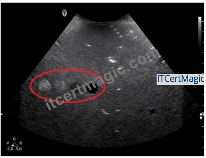
- A. Elevational
- B. Axial
- C. Contrast
- D. Lateral
Answer: B
Explanation:
The tissue-equivalent phantom image with the red oval indicates an area where axial resolution can be evaluated. Axial resolution refers to the ability to distinguish between two structures that are close together along the axis of the ultrasound beam. It is determined by the spatial pulse length (SPL) of the ultrasound wave. In phantoms, this is typically tested by observing the ability to separate closely spaced targets along the beam's path.
Reference:
ARDMS Sonography Principles & Instrumentation Guidelines
Hedrick WR, Hykes DL, Starchman DE. Ultrasound Physics and Instrumentation. 4th ed. Philadelphia, PA: Elsevier Saunders; 2005.
NEW QUESTION # 22
What is the effect of an increased aperture in a linear array transducer?
- A. Improved axial resolution
- B. Deeper focus
- C. Decreased temporal resolution
- D. Shorter near-field length
Answer: B
Explanation:
The aperture of a transducer is the active area that emits and receives the ultrasound waves. In a linear array transducer, increasing the aperture (using more elements for transmission and reception) results in a deeper focus because the beam is more tightly focused over a longer distance. This improves lateral resolution at greater depths, as the ultrasound beam maintains a narrower width for a longer distance. It allows for better imaging of deeper structures without sacrificing resolution.
Reference:
American Registry for Diagnostic Medical Sonography (ARDMS). Sonography Principles and Instrumentation (SPI) Examination Review Guide.
NEW QUESTION # 23
How can the spectral Doppler mirroring seen in this image be eliminated?
- A. Increase dynamic range.
- B. Increase pulse repetition frequency (PRF).
- C. Decrease Doppler gain.
- D. Decrease wall filter.
Answer: C
Explanation:
Spectral Doppler mirroring, also known as crosstalk, occurs when the Doppler signal appears on both sides of the baseline. This can be caused by excessively high Doppler gain, which amplifies the signal and creates artificial mirror images. Decreasing the Doppler gain reduces the signal amplitude, thereby minimizing the mirroring artifact.
Reference:
ARDMS Sonography Principles and Instrumentation guidelines
Hoskins, P. R., Thrush, A., Martin, K., & Whittingham, T. A. (2010). Diagnostic Ultrasound: Physics and Equipment.
NEW QUESTION # 24
What produces increased attenuation within soft tissue?
- A. Lower intensity of the ultrasound beam
- B. Higher frequency of the ultrasound beam
- C. Higher intensity of the ultrasound beam
- D. Lower frequency of the ultrasound beam
Answer: B
Explanation:
Attenuation refers to the reduction in the intensity of the ultrasound beam as it travels through tissue. Higher frequency ultrasound beams experience more attenuation because they are absorbed and scattered more than lower frequency beams. This is due to the fact that higher frequency waves have shorter wavelengths and interact more with the small particles in tissues, causing greater energy loss.
Reference: ARDMS Sonography Principles and Instrumentation, Chapter on Ultrasound Physics and Instrumentation.
NEW QUESTION # 25
Which unfocused transducer will have the greatest divergence?
- A. 6 mm aperture, 4 MHz
- B. 4 mm aperture, 6 MHz
- C. 4 mm aperture, 4 MHz
- D. 6 mm aperture, 6 MHz
Answer: C
Explanation:
Transducer beam divergence is influenced by the aperture size and frequency. A smaller aperture and lower frequency result in greater beam divergence. Among the given options, the transducer with a 4 mm aperture and 4 MHz frequency will have the greatest divergence. This is because the smaller aperture size contributes to a wider beam spread, and the lower frequency also increases the divergence compared to higher frequencies.
Reference:
ARDMS Sonography Principles and Instrumentation guidelines
Kremkau, F. W. (2015). Diagnostic Ultrasound: Principles and Instruments. Elsevier.
NEW QUESTION # 26
Which type of display process rescans only the region of interest and improves resolution?
- A. Frequency compounding
- B. Spatial compounding
- C. Write magnification
- D. Read magnification
Answer: C
Explanation:
Write magnification, or pre-processing zoom, involves rescanning the region of interest (ROI) with more scan lines, thus acquiring new data for that specific area. This process increases the spatial resolution of the image in the magnified area because it gathers more detailed data by adjusting the scan parameters, resulting in improved image quality. This is different from read magnification (post-processing zoom), which simply enlarges the existing image data without increasing resolution.
Reference:
ARDMS Sonography Principles & Instrumentation Guidelines
Hedrick WR, Hykes DL, Starchman DE. Ultrasound Physics and Instrumentation. 4th ed. Philadelphia, PA: Elsevier Saunders; 2005.
NEW QUESTION # 27
What does changing the displayed depth control directly affect?
- A. Pulse repetition frequency
- B. Pulse duration
- C. Transducer transmit frequency
- D. Spatial pulse length
Answer: A
Explanation:
Changing the displayed depth control directly affects the pulse repetition frequency (PRF). When the depth setting is increased, the ultrasound system needs more time to send and receive echoes from deeper structures, resulting in a lower PRF. Conversely, decreasing the depth allows for a higher PRF since the time required for the sound waves to travel to and from the structures is shorter. PRF is crucial for determining the maximum detectable velocity in Doppler ultrasound without aliasing. Reference:
ARDMS Sonography Principles and Instrumentation guidelines
"Understanding Ultrasound Physics" by Sidney K. Edelman
NEW QUESTION # 28
Which statement characterizes the primary difference between image A and image B?
- A. Image A demonstrates a better axial resolution.
- B. Image A demonstrates a wider scale of contrast.
- C. Image A demonstrates a shallower field of view.
- D. Image A demonstrates a lower overall gain setting.
Answer: D
Explanation:
The primary difference between Image A and Image B is the overall gain setting. Gain controls the amplification of the received echoes. A lower gain setting results in a darker image with less overall brightness, which is evident in Image A compared to Image B. Image B appears brighter, indicating a higher gain setting that amplifies the echoes more, making the structures appear more prominently.
Reference:
ARDMS Sonography Principles and Instrumentation guidelines
Hedrick, W. R., Hykes, D. L., & Starchman, D. E. (2005). Ultrasound Physics and Instrumentation.
NEW QUESTION # 29
The calipers in this image measure which performance characteristic of a system?
- A. Lateral resolution
- B. Depth measurement accuracy
- C. Dynamic range
- D. Axial resolution
Answer: B
Explanation:
The calipers shown in the image are used to measure the depth of structures within the ultrasound image. This performance characteristic, known as depth measurement accuracy, assesses how accurately the ultrasound system can measure the distance from the transducer to a specific point within the body. Accurate depth measurements are crucial for diagnostic purposes, ensuring that anatomical and pathological structures are correctly identified and evaluated.
Reference:
American Registry for Diagnostic Medical Sonography (ARDMS) Sonography Principles and Instrumentation study materials.
Textbook of Diagnostic Sonography by Hagen-Ansert, S. L. (latest edition).
NEW QUESTION # 30
At which angle to blood flow would the maximum Doppler shift occur?
- A. 60 degrees
- B. 30 degrees
- C. 0 degrees
- D. 90 degrees
Answer: C
Explanation:
The Doppler shift is highest when the angle between the ultrasound beam and the direction of blood flow is 0 degrees. This is because the cosine of 0 degrees is 1, maximizing the Doppler frequency shift. As the angle increases towards 90 degrees, the cosine value decreases, reducing the Doppler shift.
Reference:
ARDMS Sonography Principles and Instrumentation guidelines
Hoskins, P. R., Thrush, A., Martin, K., & Whittingham, T. A. (2010). Diagnostic Ultrasound: Physics and Equipment.
NEW QUESTION # 31
What is the relationship between overall gain and image brightness?
- A. The higher the overall gain, the brighter the image
- B. There is no relationship between overall gain and image brightness
- C. The lower the overall gain, the brighter the image
- D. The higher the overall gain, the darker the image
Answer: A
Explanation:
Overall gain in ultrasound refers to the amplification of all the received echo signals. Increasing the overall gain amplifies the signals, making the entire image brighter. Conversely, decreasing the overall gain reduces the signal amplification, resulting in a darker image. Overall gain adjustment affects the entire image uniformly, unlike time gain compensation (TGC), which adjusts the gain at different depths independently.
Reference:
ARDMS Sonography Principles and Instrumentation (SPI) Exam Study Guide
"Diagnostic Ultrasound: Principles and Instruments" by Frederick W. Kremkau
NEW QUESTION # 32
What does damping in a pulsed-wave transducer affect?
- A. Pulse duration
- B. Beam penetration
- C. Pulse repetition frequency
- D. Beam focus
Answer: A
Explanation:
Damping in a pulsed-wave transducer primarily affects the pulse duration. Damping refers to the process of reducing the vibration of the transducer crystal after the initial excitation. When damping is applied, it shortens the length of the ultrasound pulse by quickly reducing the vibration. This results in a shorter pulse duration, which is important for improving axial resolution. Damping does not directly affect the pulse repetition frequency, beam focus, or beam penetration, although it can have indirect effects on image quality and resolution. Reference:
ARDMS Sonography Principles and Instrumentation guidelines
"Diagnostic Ultrasound: Principles and Instruments" by Frederick W. Kremkau
NEW QUESTION # 33
Which adjustment can maintain the same frame rate when the depth is increased?
- A. Increase number of focal zones
- B. Decrease persistence
- C. Decrease image width
- D. Increase frequency
Answer: C
Explanation:
When the depth of imaging is increased, the time it takes for the ultrasound pulses to travel to and from the deeper structures also increases, which can reduce the frame rate. To maintain the same frame rate, one effective adjustment is to decrease the image width. Narrowing the image width reduces the number of scan lines required to create each frame, allowing the system to maintain a higher frame rate despite the increased depth.
Reference:
ARDMS Sonography Principles and Instrumentation guidelines
Kremkau, F. W. (2015). Diagnostic Ultrasound: Principles and Instruments.
NEW QUESTION # 34
......
Genuine SPI Exam Dumps Free Demo Valid QA's: https://www.itcertmagic.com/ARDMS/real-SPI-exam-prep-dumps.html
Printable & Easy to Use ARDMS SPI SPI Dumps 100% Same Q&A In Your Real Exam: https://drive.google.com/open?id=1fDn70sa-zPWmySvvo5kJt_UIFgUmK38d